Equipment: iEEG electrodes
Several forms of iEEG exist, with the defining feature of each being the type of electrode used and its method of placement. The measurement of electrical activity in the brain using subdural grid or strip electrodes is called electrocorticography (ECoG). Studies using depth electrodes in data collection are referred to as stereotactic EEG (sEEG) (Parvisi & Kastner, 2018). The following table provides an overview of the various electrodes that may be used in iEEG studies.
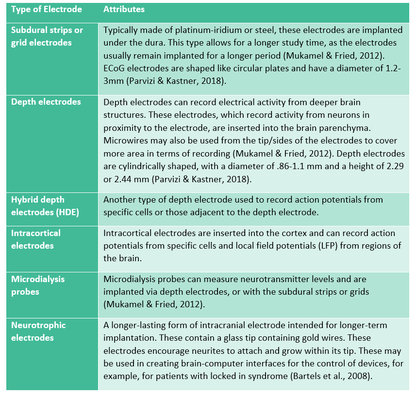
A combination of different types of electrodes may be used in the same patient in many cases. It is additionally possible to stimulate various regions of the brain with the use of some types of electrodes during awake brain surgery for functional brain mapping (Mukamel & Fried, 2012). Deep brain stimulation (DBS) involves the permanent implantation of electrodes for chronic neurological conditions like Parkinson’s disease. As these electrodes are permanently implanted, they may be used in iEEG recording for longer periods of time. Neurotrophic electrodes may be implanted in the long term to form a brain-computer interface which permits the control of external devices for patients with locked-in syndrome or other forms of paralysis.
Channel outputs use a system of reference montage in which the difference in electrode output is compared to either a nearby electrode (bipolar reference) or one reference electrode (Stolk et al., 2018). Knowledge of the placement of the reference electrode, if this scheme is employed, is important, as placement near certain musculature can potentially introduce noise into the data (Lachaux et al., 2003; Nejedly et al. 2018). This is less of a problem with bipolar referencing schemes (Kovach et al., 2011; Jerbi et al., 2009). However, it does pose the risk of not being able to measure fluctuations that result similarly in both electrodes in the bipolar reference and may be subject to “travelling waves”, or the bleed over of signal from neighboring electrodes (Lachaux et al., 2003). Therefore, referencing information, including the exact placement of reference electrodes, should also be reported as part of the dataset’s metadata because of their implications for the data (Holdgraf et al., 2019). The numbering of the grid and strip electrodes also requires that the research have access to notes acquired under surgery and data acquisition (Stolk et al., 2018).
Researchers at UiO utilize two data acquisition systems for iEEG amplification and data collection: the Micromed system, which is an audio-video iEEG monitoring system for sEEG, and the Neuralynx System.
Usually two computers are used in iEEG recording. One computer presents the stimuli and monitor behavior, while the second records the iEEG. These functions are not ordinarily combined into a single computer, because each requires precise timing, and it is difficult to coordinate multiple real-time streams of events on a single computer. For later analysis, stimulus presentation and responses must be recorded along with the iEEG signal. For this purpose, both the A/D box and the stimulus computer are connected to a trigger box where event codes from the stimulus computer are integrated with the iEEG signal. These event codes are later used to extract time-locked epochs from the iEEG data.